
Inside the MRI: What Actually Happens During an MRI Scan?
Magnetic Resonance Imaging (MRI) is an important diagnostic tool in modern healthcare. It allows physicians to examine internal structures of the body, including the brain, spine, muscles, joints, and organs, without using ionizing radiation. However, for many patients, the process can seem unfamiliar. They enter a scanner, hear loud tapping and knocking sounds, and are asked to remain still while the examination takes place.
This often raises several questions: how long does an MRI take, why does the machine make so much noise, and what happens to the images after the scan is complete?
Understanding the process can help patients feel more comfortable and appreciate the technical work involved in medical imaging. It also provides an insight into the responsibilities of MRI technologists, who play an important role in patient care and diagnostic imaging.
What Happens Before an MRI Scan?
An MRI examination begins before the patient enters the scanning room. The healthcare team first completes several preparation and safety procedures to ensure that the examination can be performed appropriately.
Patient Screening and Safety Checks
Patients are generally asked to complete a screening questionnaire covering their medical history, previous surgeries, implanted devices, and other relevant health information.
This step is particularly important because MRI scanners use powerful magnetic fields. Certain metal objects and medical devices can present safety risks or interfere with image quality.
Patients should inform the healthcare team about:
- Pacemakers, implants, surgical clips, and other implanted medical devices.
- Previous surgeries or metallic fragments in the body.
- Pregnancy or the possibility of pregnancy.
- Kidney conditions or relevant allergies, particularly when contrast material may be used.
- Tattoos, medication patches, or other items that may require additional screening.
Not every implant prevents someone from having an MRI. Some devices are classified as MR Safe or MR Conditional, meaning they can be used under specified conditions. The MRI team must verify the device and follow the applicable safety requirements.
Getting Ready for the Scanner
Depending on the examination, patients may be asked to change into a hospital gown and remove jewelry, watches, phones, keys, and other personal belongings containing metal.
The technologist then explains the procedure and helps the patient understand what to expect. Earplugs or headphones are usually provided because MRI scanners produce loud sounds during image acquisition.
What Actually Happens Inside an MRI Machine?
MRI technology works through the interaction of magnetic fields, radiofrequency energy, and computer processing. Unlike conventional X-ray imaging, it does not rely on ionizing radiation.
The human body contains large amounts of water and fat, both of which contain hydrogen atoms. MRI scanners use signals associated with hydrogen nuclei to create detailed images of internal tissues.
Here is what happens during the examination.
1. The Magnetic Field Aligns Hydrogen Protons
The MRI scanner produces a strong magnetic field that influences the behaviour of hydrogen protons within the body. This creates a measurable magnetic response that forms the basis of MRI imaging.
2. Radiofrequency Pulses Interact With the Protons
The machine sends radiofrequency pulses into the body. These pulses change the magnetic state of the protons, which subsequently produce signals as they return towards their equilibrium state.
Different tissues produce different signal characteristics. These differences help distinguish structures and abnormalities within the body.
3. The Scanner Collects Signals
Special receiver coils detect the signals produced during the examination. Magnetic field gradients help identify where the signals originate, allowing the scanner to collect spatial information from the selected body area.
4. Computer Processing Creates Images
The collected information is processed by a computer and reconstructed into detailed images. These images can represent different tissue characteristics and anatomical sections, giving radiologists information that helps them assess the area being examined.
The complete process involves several imaging sequences rather than one continuous photograph. Each sequence is designed to provide specific information about the tissues.
Why Does an MRI Machine Make Loud Noises?
One of the most noticeable parts of an MRI examination is the sound produced by the scanner. Patients commonly hear clicking, knocking, tapping, and rhythmic thumping noises.
These sounds are normal and are mainly produced by the gradient coils switching rapidly during image acquisition. These coils change the magnetic field in controlled ways to help the system determine the location of the signals being collected.
Different imaging sequences produce different sound patterns because the scanner changes how it operates throughout the examination.
Patients are provided with hearing protection to reduce exposure to scanner noise. The MRI technologist also monitors the examination and maintains communication with the patient through an intercom or other communication system.
How Long Does an MRI Take?
One of the most common questions patients ask is how long does an MRI take. The answer depends on the body part being examined, the number of imaging sequences required, the equipment, and the clinical purpose of the examination.
Many routine MRI examinations take approximately 30 to 60 minutes. Some scans can be completed in less time, while more detailed examinations may take longer.
For example, an examination involving multiple anatomical areas or additional imaging sequences may require more time than a focused examination of a single joint.
The overall appointment can also take longer than the actual scanning process because patients must complete safety screening, change clothing when required, receive positioning instructions, and prepare for the examination.
If contrast material is required, additional preparation and image acquisition may also be necessary.
Why Do MRIs Take So Long?
Patients sometimes wonder why do MRIs take so long when the actual procedure appears to involve simply lying inside a machine.
The explanation is that MRI examinations involve several stages of data collection. Each stage provides different information needed to produce images suitable for clinical interpretation.
Multiple Imaging Sequences
An MRI examination may include several sequences, each designed to highlight different tissue characteristics. For example, T1-weighted and T2-weighted images provide different types of anatomical information.
The scanner must collect the required data for each sequence before the final examination is complete.
High Image Resolution
Detailed images require accurate signal collection and processing. Depending on the examination, obtaining the required resolution and coverage can take additional time.
The number of images, selected imaging parameters, and area being examined all influence the duration.
Patient Movement
Remaining still is important during an MRI examination. Even small movements can affect image quality and create motion artefacts.
When an image is unclear because of movement, the technologist may need to repeat a particular sequence. This can extend the examination.
Use of Contrast Material
Some MRI examinations involve gadolinium-based contrast agents to provide additional information about certain tissues and structures.
When contrast is required, the examination may include additional preparation, an injection, and further image sequences. Contrast is not necessary for every MRI examination.
Different Clinical Requirements
Not every MRI examination has the same purpose. A brain MRI, spinal MRI, cardiac MRI, and joint MRI involve different anatomical structures and imaging requirements.
The radiologist’s clinical question and the approved examination protocol determine the type and amount of information that needs to be collected.
What Happens After an MRI Scan?
Once the examination is complete, the patient is helped off the scanning table. If an IV line was used for contrast administration, it is generally removed when no longer needed.
The technologist may also review the images for technical quality and determine whether additional pictures are required before the patient leaves.
However, completing the scan does not mean the diagnostic results are immediately available.
The collected images are reviewed by a radiologist, a physician trained in interpreting medical imaging examinations. The radiologist evaluates the images, identifies relevant findings, and prepares a report for the referring healthcare professional.
The referring physician then discusses the findings with the patient and explains any recommended follow-up or treatment decisions.
How Long Does It Take to Get MRI Results?
Another common concern is how long does it take to get MRI results after leaving the imaging center.
There is no universal reporting timeframe. The time depends on the healthcare facility, the radiologist’s workload, the complexity of the examination, and whether the case requires urgent review.
Some reports may be completed relatively quickly, while routine examinations at busy facilities can take longer. Patients should ask their imaging facility or referring physician about the expected reporting schedule.
Understanding MRI Scanning Result Time
The term MRI scanning result time can refer to two different stages: the time required to capture the images and the time needed for a radiologist to interpret them.
These are separate processes.
The scanning appointment may be completed in under an hour, but reviewing the images requires additional professional assessment. A radiologist may need to examine multiple sequences, compare relevant previous studies, and prepare a written report.
A delay in receiving results does not, by itself, indicate whether the findings are positive or negative. Patients should contact the referring healthcare professional or imaging facility if the expected reporting period has passed.
What Is the Role of an MRI Technologist During the Examination?
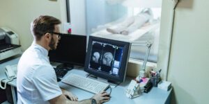
MRI technologists are responsible for important technical and patient-care activities throughout the examination. Their work helps ensure that imaging procedures follow established protocols and safety requirements.
Their responsibilities commonly include:
- Reviewing patient information and completing MRI safety screening.
- Preparing and positioning patients for the examination.
- Operating MRI equipment according to approved protocols.
- Selecting appropriate imaging parameters within their professional responsibilities.
- Monitoring patients and maintaining communication during scanning.
- Checking image quality and identifying technical issues.
- Following equipment safety and infection-control procedures.
MRI technologists must understand anatomy, magnetic resonance principles, imaging procedures, and patient safety. They also need effective communication skills to help patients understand the procedure and remain comfortable during scanning.
While MRI technologists acquire the images and assess their technical quality, the radiologist is responsible for interpreting the examination and preparing the diagnostic report.
Explore MRI Technology Education With Us
The processes involved in an MRI examination demonstrate how healthcare depends on both advanced equipment and trained professionals. Understanding magnetic resonance principles, patient positioning, image acquisition, and safety procedures forms an important part of education in this specialized field.
We offer a Magnetic Resonance Imaging (MRI) Technologist Diploma Program for individuals interested in medical imaging education.
Students considering this professional pathway can learn more about the MRI Technologist program, including its educational approach, program information, and admission requirements.
Choosing a healthcare education program requires careful consideration of academic preparation, practical learning opportunities, and the professional requirements associated with the intended career. Prospective students should review the current program information and discuss their educational goals with the admissions team.
Interested in studying MRI technology? Visit Stellar Career College to learn more about the MRI Technologist Diploma Program and schedule an orientation.
Frequently Asked Questions
1. How long does an average MRI scan take?
Many MRI examinations take approximately 30 to 60 minutes. However, the duration depends on the body part being examined, the required imaging sequences, and whether additional procedures are necessary.
2. Why do MRI scans require patients to remain still?
Movement can create motion artefacts that distort images and make anatomical details difficult to assess. Remaining still helps produce clear images for radiologist interpretation.
3. Do all MRI scans require contrast injections?
No. Many MRI examinations are performed without contrast material. Gadolinium-based contrast agents are used when additional imaging information is clinically needed.
4. How long does it take to get MRI results?
The reporting time varies between healthcare facilities. A radiologist must review the images and prepare a report before the referring healthcare professional discusses the findings with the patient.
5. Can an MRI technologist explain the scan results?
MRI technologists can explain the examination process and provide instructions about the procedure. However, interpreting diagnostic findings and communicating the medical conclusions are responsibilities of the radiologist and referring healthcare professional.
6. Is an MRI scan painful?
Most MRI examinations are painless. However, some patients may find it uncomfortable to remain still or experience anxiety because of the enclosed scanner environment. Patients should communicate any discomfort or concerns to the healthcare team.
Related Blogs

Inside the MRI: What Actually Happens During an MRI Scan?
Learn what happens inside an MRI scanner, how long scans take, why they take time, and when to expect MRI...
The History of the MRI: Its Development and Growing Importance in Healthcare
Learn about the history of MRI, its development, and its growing importance in modern healthcare and medical imaging.

Why Is Chicago Called the Windy City? A Fascinating History for International Students
Discover why Chicago is called the Windy City, its fascinating history, student life, and healthcare education opportunities.